


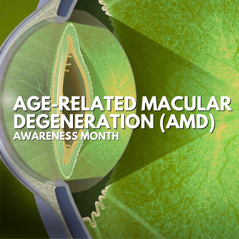
Age-Related Macular Degeneration
Age-Related Macular Degeneration (AMD): What It Is, What to Watch For, and How We Monitor It
Age-related macular degeneration (AMD) is an eye condition that affects the macula—the small, central area of the retina responsible for sharp, detailed vision needed for reading, driving, and recognizing faces. AMD can gradually blur or distort central vision, and in more advanced cases may create a dark or missing spot in the center of what you see.
Dry AMD vs. Wet AMD: What’s the Difference?
AMD generally falls into two categories:
Dry AMD (non-neovascular AMD): The more common form. It’s often associated with drusen (tiny yellow deposits under the retina) and gradual changes to the retinal pigment layer. Many people with early dry AMD have few or no noticeable symptoms, which is why routine eye exams are so important.
Wet AMD (neovascular AMD): A less common but more aggressive form of late AMD. Wet AMD happens when abnormal blood vessels grow under the retina and can leak fluid or blood, leading to faster vision changes and scarring if not treated promptly.
Importantly: any stage of dry AMD can progress to wet AMD, so ongoing monitoring matters.
Common Symptoms of AMD
Contact your eye doctor promptly if you notice:
Wavy or distorted lines (doors, blinds, text lines)
Blurry or “smudged” central vision
A dark, gray, or missing spot in the center of your vision
Trouble reading or needing brighter light than usual
Because AMD can begin quietly, you don’t want to wait for symptoms before being evaluated.
Risk Factors (and what you can control)
Age and family history play a big role, but there are modifiable factors too. Smoking is one of the most significant—smokers are about twice as likely to develop AMD compared with non-smokers, and quitting can help reduce risk.
How We Find and Monitor AMD in Our Office:
AMD care is all about two goals:
catching changes early, and
tracking the macula over time so we can act quickly if AMD progresses.
Here are the main ways we do that:
1) Dilated retinal exam
During a comprehensive exam, we use dilating drops so the doctor can get a wide view of the retina and macula. This allows us to look directly for signs like drusen, pigment changes, and other macular findings that may suggest early or progressing AMD.
2) Retinal photography (fundus photos)
A retinal camera captures detailed images of the back of your eye. These photos are incredibly helpful because they:
document what your macula looks like today,
create a baseline for comparison,
and help us detect subtle changes over time.
3) OCT Scanning (Optical Coherence Tomography)
OCT is one of the most valuable tools we use for AMD. It’s a quick, non-contact scan that uses light waves to create cross-section (“slice”) images of the retina—almost like an “optical ultrasound,” but with light instead of sound.
Why OCT matters for AMD:
It helps us see and measure the retina’s layers in high detail.
It can reveal early structural changes linked to dry AMD.
Most importantly, OCT can detect signs of wet AMD activity, like retinal swelling and fluid, which often need urgent treatment to help protect vision.
In other words: OCT helps us move from “I think things look stable” to “We can see exactly what’s happening—layer by layer.”
4) Additional testing (when appropriate)
Depending on what we see, we may recommend additional imaging or refer you to a retina specialist for specialized tests that look more closely at blood vessel changes associated with wet AMD.
How AMD Is Managed:
Treatment depends on the type and stage:
Lifestyle and risk reduction: Especially smoking cessation and overall cardiovascular health support.
AREDS2 supplements (for specific patients): For people with intermediate AMD (or advanced AMD in one eye), the AREDS2 formulation can help slow progression. These supplements aren’t for everyone, so it’s important to use them only when recommended based on exam findings.
Wet AMD treatment: Wet AMD is often treated with medications delivered by injection to reduce abnormal vessel activity and fluid leakage. Fast detection and referral are key because wet AMD can change quickly.
The bottom line:
AMD is common, often silent early on, and highly dependent on early detection + consistent monitoring. The good news: with tools like retinal photography and OCT, we can catch changes sooner, track progression more precisely, and act quickly if wet AMD develops.
Ready to protect your sight?
If you’re over 50, have a family history of macular degeneration, notice any new visual distortion, or simply want peace of mind, we’d love to help. Schedule a comprehensive eye exam at InFocus Family Eyecare and Optical Boutique so we can evaluate your macula, establish a baseline, and monitor for changes over time. Early care makes a real difference
References:
National Eye Institute (NIH) — Age-Related Macular Degeneration overview.
American Academy of Ophthalmology — Understanding Macular Degeneration (dry vs. wet, symptoms, treatment overview).
American Academy of Ophthalmology — What Is Optical Coherence Tomography (OCT)?
National Eye Institute (NIH) — AREDS/AREDS2 clinical trial summary (supplements and progression risk reduction).
American Academy of Ophthalmology — Anti-VEGF treatments (wet AMD therapy concept).
NCBI Bookshelf (NIH) — Overview: Age-related macular degeneration (general patient overview, dry vs wet).
Quick Links

All Eye
Care Services

Keep
In Touch
MAP
© 2026 InFocus Family Eyecare. All Rights Reserved. Accessibility Statement - Privacy Policy - Sitemap
Powered by: 